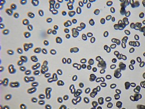
I’ve been spending a lot of time sorting out how to look at spores under a microscope. I was given a fairly basic microscope from Lidl for Xmas 2013, which at £50 is astounding value and in theory can magnify up to x1280. It’s taken a while to get up to speed and gain familiarity with the setup, lighting and focusing required. The basic problem is that mushroom spores are so small, and it’s rather tricky trying to use a cheap microscope, but the results are surprisingly good.
All the pictures when transferred to computer at maximum resolution are 640×480 pixels. A classic problem is how to work out the size of the spores in the picture. My approach is to measure the spore length in pixels of a known mushroom species with a known spore length and thereby deduce the length of a pixel. I did that with 6 species and came up with the answer of 2.6 pixels per micrometre.
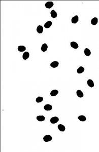
It’s quite hard to measure the length and width (in pixels) of a whole load of spores. So I wrote a Java program to do it. (Java design and programming is my day job.) It reads the jpeg, converts it into a black and white image using the best black/white cut-off level that gives a whole load of blobs with similar size, then writes the lengths and widths out to a csv file that can be opened with Excel.
This is my analysis procedure:
1. Get the best in-focus picture possible.
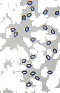
2. Edit the picture in Windows Paint, cropping to get the area with the best samples, and rubbing out touching/overlaid spores. Ideally need 20+ spores, but 10 will do.
3. Run the picture through my image anaysis program to get the length and width of all the spores. Check the black/white image produced by the program with the original image, to ensure that it looks sensible.
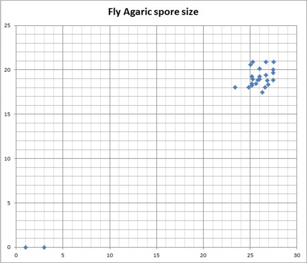
4. Divide pixel lengths and widths by 2.6. Plot the length v. width data points on a scatter chart. Eliminate any obvious outliers that are not actual spores, and calculate the mean and standard deviation. (Easy to do this in a spreadsheet.)
5. 95% of spore lengths or widths should be within (Average ± 2 Standard Deviation). So obtain:
(Average spore length – 2*SD)
(Average spore length + 2*SD)
(Average spore width – 2*SD)
(Average spore width + 2*SD)
6. Compare with the book values
Fly Agaric: step 1 step 2 step 3
Fly Agaric: step 4
| Fly Agaric steps 5,6 | spore length | spore width | |||
| min | max | min | max | ||
| Observed | pixels mean±2sd | 24 | 28 | 17 | 21 |
| Observed | pixels/2.6 mean±2sd | 9.4 | 10.7 | 6.6 | 8.2 |
| Book values | µ | 9.5 | 10.5 | 7 | 8 |
Spore Pictures for 7 species:
- Ugly Milkcap spores
- Lactarius controversus spores
- Horse Mushroom spores
- Fairy Ring Mushroom spores
- Cultivated Mushroom spores
- Blushing Bolete spores
- Fly Agaric spores
Table of results for 7 species:
| Shape | Book values | Observed values | Remarks | |
| Blushing Bolete | Book says subfusiform (somewhat spindle shaped, narrowing at both ends). Yes. | 15.5-17.5 x 5-5.5µ | 13-19.5 x 6-8µ | Observed spore length has greater range of values than book, and observed width is wider. Looking at the sample, a fairly wide range of sizes is clearly evident. Perhaps also the picture is a little fuzzy which might be exaggerating the size. |
| Cultivated Mushroom | Book says ovate (egg shaped) to subglobose (somewhat spherical). Yes. | 4-7.5 x 4-5.5µ | 6-8 x 4.5-6.5µ | Observed spores are at maximum of book values. Perhaps there are varieties of shop-bought mushrooms with somewhat different spore sizes? |
| Fly Agaric | Book says broadly ovate (egg shaped). Yes. | 9.5-10.5 x 7-8µ | 9.5-10.5 x 6.5-8µ | Spot on. |
| Horse Mushroom | Book says elliptical. Yes. | 7-8 x 4.5-5µ | 5.5-7.5 x 4.5-5.5µ | About the same. |
| Ugly Milkcap | Book elliptical; ridges tending to run across the spore, forming a fairly well-developed network. Yes, they are elliptical. And I can even see ridges on a few, but there’s no chance of spotting a network. | 7.5-8.5 x 6-7µ | 6.5-8 x 5.5-6.5µ | Observed spores are at minimum of book values. |
| Lactarius controversus | Book says oval; strong warts connected by thick ridges forming a fairly complete network. Yes, they are oval. And I’ve managed to convince myself that I can see warts. Not sure about a network, although they does seem to be some kind of ornamentation on some. | 6-7 x 5-6µ | 5.5-8 x 4.5-6µ | Observed spores are at minimum of book values. I originally thought this was Fleecy Milkcap (7.5-9.5 x 6.5-8.5µ) but based on this spore evidence it can’t be. Before seeing this evidence I was very unsure whether this was Fleecy Milkcap or Lactarius controversus. |
| Fairy Ring Mushroom | Book says pip shaped. Yes. | 8-10 x 5-6µ | 10-10.5 x 7-7µ | Observed spores are bigger than book values. However there were just 2 spores in the only good picture I had, so can’t come to conclusions with such a small sample size. |


